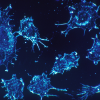
A close look at Parkinson's
Interview with
Parkinson's Disease occurs when a protein called alpha synuclein accumulates inside  certain subsets of nerve cells, causing them to die. The protein might also spread along nerve connections from an affected area to other brain regions, although this has been difficult to test without access to very large numbers of patient samples. Now McGill University researcher Alain Dagher has analysed hundreds of brain scans from Parkinson's and healthy patients to spot some key differences that single out the condition, as well as provide evidence that the disease does spread around the brain, and he spoke to Chris Smith about his findings...
Alain - Previous studies all suffered from small numbers of patients and so, they did not have the power to detect abnormalities. So the first thing we did was map the distribution of the changes in Parkinson's disease compared to healthy controls. Up to now, the only other way to do this would have been with post-mortem studies and this is indeed what pathologists have been doing including a pathologist by the name of Braak who was really one of the pioneers in proposing one of the theories that we tested in this study. This theory is that Parkinson's disease is due to the spread of a toxic agent through the brain. His theory was that the toxication spread from brain cell to brain cell. So, we know that brain cells have the ability to communicate with each other. They make connections called synapses and the theory here is that some sort of toxic product - in this case, probably a protein has the ability to be transmitted from one cell to another, a little bit like an infectious disease.
Chris - This is the basis of BSE, bovine spongiform encephalopathy and prion disease in humans like CJD. The idea is that some kind of protein accumulates and it then spreads through the brain, transmitting some kind of abnormal configuration of the protein from one area to another.
Alain - That is exactly correct. In the case of a Parkinson's disease, the protein is a normal protein called alpha synuclein which gets misfolded and it's its misfolded state that becomes toxic. Now, we can't image alpha synuclein using MRI in living humans but what we can do is ask whether the pattern of atrophy fits with a spread of a toxic agent through brain networks.
Chris - And is that what you see? If you look at the connectivity, in other words, which bits of the brain connect to each other, one would anticipate if there is some kind of transmitted phenomenon going on where some kind of pathogen goes from one brain area to another, it's going to be focused where the connections from the diseased brain area are most intensely focused or project to in the brain, you'd therefore see that as the next node in the chain. Is that what you'd see in these MRI scans?
Alain - Correct. Based on your knowledge of the normal connections of the brain and the pattern of the disease that you see, you can infer that the pattern fits with the spread of an agent through the networks.
Chris - So apart from corroborating what was a nice theory which seems to be borne out by the neuropathology, you've now got this scanned data from large numbers of people that appears to support it too, are there any other implications of what you found? Could you use your scan baseline as a template to say, stage disease or to iron out the 10, 20 per cent of people who get a wrong diagnosis of Parkinson's disease and give them a more accurate diagnosis in the future?
Alain - So, this is the hope. There are different kinds of Parkinson's disease. Different patients progress at different rates. Different patients develop different types of symptoms through the years. Because of this database, we'll include repeat evaluations of the participants. We hope to be able to track progression of the disease and eventually perhaps provide an imaging method to be able to, number one, better diagnose a disease but number two, follow the progression of the disease and eventually test therapeutic interventions. So for example, if someone develops a method to stop the spread of this protein, this imaging methodology will be useful in testing whether it's effective.
- Previous Stress and depression
- Next Good and bad parents


Comments
Add a comment