Hibernation, Ketamine and Aphantasia
This month, how animals hibernate and evidence that muscle myosin makes its own heat in the cold, brain scans to reveal how ketamine relieves resistant depression, the way the brain changes when animals build a bond, the evolution of flu outbreaks, and how aphantasia affects autobiographical memory.
In this episode

00:32 - How animals hibernate
How animals hibernate
Christopher Lewis, University of Copenhagen
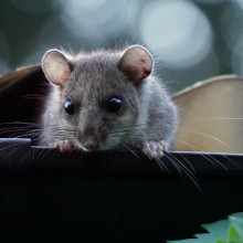
We traditionally regard us mammals as “warm blooded”, but some among our numbers have adapted to allow their body temperatures to periodically plunge, dramatically cutting their metabolic rate and thus making energy reserves last much longer. This, of course, is the basis of hibernation. But, as Christopher Lewis, working at the time at the University of Copenhagen, has found, it’s more complicated than just dialling down the temperature. Speaking with Chris Smith, he’s found that myosin in muscles can make heat just in response to becoming cold. But when very small animals - like the dormice he studied - want to hibernate, they suppress this process by putting their myosin into an altered configuration that doesn’t get hot when it’s cold. Bigger animals like bears, on the other hand, with far more energy reserves, can afford not to do this and they stay warm all the time…
Christopher - Hibernation is a unique phenomenon which certain mammals take part in every year. And it's where they will shut their body down for six months to eight months of the year in order to survive, to survive during harsh winter periods where there's little food. And really that's quite an incredible feat to be able to effectively sleep for six months. Not eat, not go to the toilet, not drink any water and actually remain healthy! The amount of energy that your body uses really shuts down.
Chris - Do all animals hibernate equally? As in if I compare a tiny mammal with say a big brown bear, is hibernation the same or are we lumping together different behaviours that look sort of similar, all under the same umbrella, but there are differences?
Christopher - Yes, there absolutely are differences. And this is something that we, we look into with the paper and, and what you find is with larger animals such as bears - in this manuscript we looked at two types of bears, brown bears and black bears - they hibernate at quite normal temperatures. Their body temperature doesn't really drop very much, maybe by a few degrees. You then have smaller mammals and they will hibernate at very, very different temperatures. Actually, their core body temperature goes from 37 down to about four degrees. And this we think is very important in actually how these animals hibernate. So this is why in this paper we looked at large hibernators but also small hibernators.
Chris - And when you say you looked at them, what did you actually study?
Christopher - We are really interested in skeletal muscle. And the reason we're interested in that is because skeletal muscle makes up a large amount of our body mass. It's a very large organ or set of organs, tissues within all mammals. And therefore it uses a lot of energy. So it's particularly important in hibernation when you're trying to reduce how much energy that you use. So what we did was we took small biopsies from each of these animals whilst they were both hibernating and active. And we were able to dissect each single muscle fibre and we were able to observe how much energy each fibre was using whilst either they were active or they were hibernating.
Chris - How do you know how much energy they use?
Christopher - We actually put them under a microscope, insulate the fibre with a fluorescent energy or ATP molecule. Because it's fluorescent, it'll make this muscle fibre glow. And therefore what we can do is we can effectively video this muscle fibre over the course of five minutes and we can watch how quickly it uses the energy by how quickly the fluorescence of the muscle fibre decreases. The quicker that the glow disappears, the more energy the muscle fibre is using.
Chris - And what's actually doing the energy consumption? Where's the energy going?
Christopher - Yeah. So what we were really interested in in this study is something called myosin. It is the motor unit which allows our muscle to contract. It effectively uses energy directly and it moves in order to allow the muscle to move. However, what has been recently discovered is that even when you're resting, so when you're sleeping for example, or hibernating, your muscle still uses energy in the same manner. However, this is very variable and the myosin conformation is able to very much dictate how much energy is being used by your muscle when you're resting. So what we were interested in, of course, was monitoring this in hibernation to see if there is a change in myosin in animals which are hibernating.
Chris - And also, can I preempt one possible other question that must have occurred to you? Yeah. Which is when you've got these big animals and small animals and the big ones stay warmer and the small ones allow themselves to get a lot colder, do they have a difference in how much energy their myosin is is burning off at the time in order to contribute to that?
Christopher - Yeah, exactly. So that was a key question for us in this, in this study, what we did is we did these experiments at different temperatures to try and mimic the kind of environment that these animals would be experiencing during hibernation. And what we found was that when we cooled down the temperature of the muscle fibre, the amount of energy that it used actually went up. And the reason why we think this is, is because it's kind of similar to shivering. When you're cold, your muscles start to shiver. And this actually uses energy to produce heat. So what we think we found in this paper is a new way which your muscle uses energy to produce heat. And what was particularly interesting in these small mammals which are very cold, whilst they hibernate, when we cooled down the hibernating muscle, it didn't increase the energy which it was using, which is almost certainly very important for its survival to allow it to maintain a cold body temperature.
Chris - And that's the key distinction, I suppose, isn't it? That means these animals that are smaller do allow themselves to get much colder. Because, what, do they stop their myosin doing this, or do they have a different form of myosin that means they're not able to produce heat in this way?
Christopher - Yeah. So they stop their myosin from doing this. The myosin undergoes changes to its structure and this stops it from using energy. And this only happens in the animals, which were cold and hibernating. So it's, yeah, as I said, it's definitely a kind of new novel way of we've, I think, discovered of how animals can produce heat via using energy in their resting muscle.
Chris - Now 1,001 other questions exist though, don't they? Because upstream of this is still whatever the trigger is that says to the animal, turn this process on or change your myosin in this way. Is it just temperature in its local sensation of the muscle or is there some sort of central trigger that the animal releases to make this happen?
Christopher - I think it's a really good question because there are so many triggers which occur both on the onset of hibernation in these animals, but also before hibernation. So for example, before they go into hibernation, they start to eat a lot of high fat foods to effectively store energy that actually can affect the signaling pathways within the muscle and the fat. And this could actually potentially be one of the triggers which is telling the myosin to start to behave differently in preparation for hibernation. Another way it might be via the brain appetite is controlled for the brain. So it is possible there's a crosstalk between the, the kind of central nervous system and the muscle for the onset of hibernation to make myosin enter this kind of a different state during hibernation. But that is what we don't know and what we would very much like to know because we think it'd be very important.

07:57 - How ketamine treats resistant depression
How ketamine treats resistant depression
Alan Anticevic, Yale
Depression is very common. Estimates are that as many as one in six people are affected, and up to a third of those might have depression termed “treatment resistant”, which fails to respond to the current gold-standard therapies. In recent years this has led researchers to explore the “drug space” more widely to look for compounds that might bring relief to those with these harder-to-manage conditions. Hallucinogens and ecstasy have shown some benefits, alongside the anaesthetic agent ketamine, which appears to be very effective short term, although neuroscientists aren’t sure why. To shed some light on what it might be doing, and speaking with Chris Smith, Alan Anticevic, at Yale, has been brain scanning healthy volunteers both on and off the drug…
Alan - Fundamentally, we still don't understand the abnormalities in the brain of people who suffer from those symptoms. And ultimately that limits how we're developing drugs for specific mechanisms that may be altered. And so this programme of research is designed to try to understand how drugs that are potential therapies affect the human brain.
Chris - Which drugs specifically?
Alan - We have studied a drug that's called ketamine. A substance that binds to a receptor called the NMDA receptor. And at a particular dose, this drug has been shown to be a potentially revolutionary medicine for people suffering from depression. Ironically, we still don't fully understand how it works. And so this study was designed to interrogate the effects of ketamine in healthy adults and to understand the patterns of brain activity that arise while people are given a dose of ketamine known to be therapeutic for depression.
Chris - Because it's an anesthetic agent in high doses, isn't it? We put people to sleep with ketamine or we use it when we do certain procedures, because It also makes them forget what's happening to them...
Alan - Indeed it is. And what's fascinating about ketamine, it has what's called a dose dependent effect on the brain. Which means that at different doses of the drug you can expect it to do different things. So in fact, at a higher dose it becomes an anesthetic. Yet at a far, far lower dose, the drug is able to change communication between brain cells in such a way that very rapidly affects a person's mood who has been suffering from depressive symptoms. And here we are studying that low dose pattern, not the anesthetic dose pattern.
Chris - And how long does the therapeutic effect last? And do we have any insights into who derives benefit? If you give this to somebody with what sort of certainty can you say they're going to feel better?
Alan - That's the billion dollar question. We as a field don't yet have a full understanding of who is most likely to benefit. And the effect of this drug is relatively short acting. So you will have a rapid antidepressant response in some people and that response will not last. And there are complicated reasons why that's the case. So it is our job and one of the reasons why we executed this study is to understand how ketamine affects the brains of different people and to identify whether there are really different patterns in the human brain of the effects of ketamine, which could give us a lens onto its potentially diverse therapeutic potential for different people.
Chris - So how did you actually go about testing how it works?
Alan - We asked 40 healthy volunteers to participate in a brain scan during which they were administered either an infusion of placebo - saline - or ketamine. We imaged their brain using a technique that's called functional magnetic resonance imaging, fMRI, for short to measure activity over time. This allows us then to analyse the patterns of the functional change that is induced by ketamine in different people.
Chris - So you've got a sort of before and after you can see what the connectivity map is, what bits of brain are talking to what other bits of brain, then you put the ketamine in and you can see how that shifts?
Alan - That's exactly right. So we, and we can do that for every single person in this study and we can understand if there is a different pattern of that shift across different people. And because the effect of ketamine is relatively strong at that dose, we could identify these different signatures in a sample of 40 people.
Chris - Do you see a consistent signature or do you see a mixture of signals that differ between individuals? Because, obviously, the pharmacologists - the people who make drugs - would love you to say, we see the same thing happening in these parts of the brain in everybody and then they know exactly where to target with the next wonder drug. But if you see a mixture of things happening, it's much harder to argue a case for exactly what's going on. So which is it?
Alan - Yeah, so that's a brilliant question. Exactly to your point, there is this assumption or, or in a lot of cases the hope that if you give a drug that is therapeutic, it's going to do the same thing in every person. Right? And therefore its biology is what we would call clean. But what we have found, we used a procedure, a mathematical procedure that allows us to test if there are more than one dimension or more than one way that people respond to ketamine. And we have found that in fact there are more than one way and two different patterns emerged as statistically significant, which we have then studied and interrogated further in this analysis. So the implication is in fact ketamine doesn't do the same thing to every person.
Chris - One study I read suggested that about two thirds of people with chronic depression that's been really hard to treat, get some benefit from being treated with ketamine. So if you take your findings, do you think that you can spot some kind of signature in the brain activity that would give you a prediction as to who is likely if they were to take ketamine to get benefit so we know who to focus on?
Alan - Yeah, exactly. So using these kinds of approaches, we can can hone in on a pattern of the effects of ketamine that may be therapeutically predictive and isolate it from patterns that are not therapeutically predictive. And so again, it goes back to your, one of your original questions, which is if the drug is inducing the same response or very similar responses in everybody, then you're working with the same features. In other words, the same effects in everybody. But if it is different, that individual variation is critical to map and that will improve the predictive models because we're focusing on the right kind of variation, the variation that is therapeutically predictive.

15:10 - 4d brain map of pair bonding
4d brain map of pair bonding
Some animals pair for life. These bonds are crucial for parental care, defence of territories and other resources, and well-being. We humans also form long term, special relationships with lovers and close friends. But what’s at the neurological heart of this process? Speaking with Chris Smith, Steven Phelps, at the University of Texas at Austin, wondered the same thing and set about developing a way to track in four dimensions - both throughout the brain and across time - how different regions of the nervous system respond to the building of a bond…
Steven - We wanted to make a map of the brain as it forms a bond. And to do that, we turn to the prairie vole, which is a rodent that lives in the Midwest, is famous for forming bonds between males and females as they mate repeatedly over the course of a day. And we do have a list of brain regions roughly 18 or so that were thought to be involved in pair bonding. But those were all regions that were chosen piecemeal based on bits of literature that we knew. And no one had taken a really systematic approach at looking at the brain in its entirety as a bond as being formed. And that that's really what was new about our experiment.
Chris - And that's what you did? You were actually able to document the entire brain as these bonds form?
Steven - Exactly. What we did was to create a three-dimensional model of the prairie vole brain that had boundaries between different brain regions built into the model so that we could take images of prairie vole brains as bonds were forming, and identify markers that are expressed in cells as they're active, visualise those markers and then map them back onto this brain. And with this three dimensional model, we could essentially count all the active neurons across the entire brain and assign them an address, say they belong to this brain region or to that brain region from animals at different key points in time as a bond is being formed.
Chris - How do you know that because you are registering activity in different brain regions when these bonds are forming, how do you know that that activity is linked exclusively to the bonding that's going on and not to the fact the animals are smelling each other, dancing around each other? There's lots of other things going on that could be mixed up with the bonding, but you don't know that they're actually part of the bonding process. They're just going on at the same time.
Steven - Well, of course, that's always a potential confound. We dealt with that in part through our experimental design. So we didn't look just at animals that were mating and forming bonds. We also looked at animals housed together as siblings. Mating is an essential part of bond formation in the prairie vole, as it is in humans and other species. Copulation initiates mechanisms of bond formation and repeated copulation that helps us form bonds. One thing that we really can't distinguish is brain activity related to sexual behaviour and brain activity related to bonding per se. And in the prairie vole they're kind of one and the same because the sexual behaviour leads to a bond. So we didn't feel like it was essential to tease those apart, but it would be really interesting to repeat our study using another species that has similar sexual behaviour but doesn't translate that sexual behaviour into a bond. And, and indeed that's one of the things we want to do next.
Chris - I was going to ask that precise question because there's another vole which doesn't form long-term bonds, isn't there? Mm-Hmm. <affirmative>. And so one could ask, do we see some of these areas you've identified as essential and part of the bonding process not lighting up in the brain of the animal that doesn't form these long-term bonds?
Steven - That's exactly what we would expect to see. And our interpretation of our findings is that we see different circuits in the brain lighting up at different points in time as a bond forms and early before a bond has really formed, but after a lot of mating has taken place. The circuits we see are circuits that are known to be involved in sexual behaviour. Later, though we see other kinds of circuits showing up that are not known to be involved in sexual behaviour per se. And we think these are are likely places where the experience of sex gets translated into some enduring bond. And these are areas I would expect not to show up in as other promiscuous species that don't form bonds like the meadow vole or the montane vole, famously.
Chris - And that's the key point here, which is have you found any additional areas which had been missed by previous attempts to, to study bonding, which also clearly fit in with our understanding of the neuro anatomy, the connectivity, and therefore the ultimate physiology, the behaviour of the animals, when they bond or don't bond?
Steven - Yes. We find in particular regions of the brain that are part of a network of emotionally related brain regions that we call the extended amygdala. And these are regions that receive lots of kind of complex sensory information. They're often involved in emotional regulation, often directed at specific individuals. They seem to encode aspects of the identity of our social partners. And we find some of these regions which project to hormonal centres that govern hormonal release. We find activity in these brain regions lights up in, in places not previously linked to bonding, but which make a lot of sense in terms of our understanding of the anatomy of the brain and its functions.
Chris - The amygdala is also linked to fear, isn't it? So is it possible that people are, are frightened to leave their partner or your voles are frightened to, to break up of what might be the consequence of of non-monogamy <laugh>?
Steven - That is a great question. The, the amygdala is linked to fear, but it's actually, when we think of fearfulness and the amygdala, we're usually talking about a really specific region of the amygdala called the central amygdala. And that doesn't really show up so much in our study. It's really other regions of the amygdala that have been implicated in social reward, in constellation behaviour and a variety of things that are showing up. It's probably not a change in fearfulness per se.
Chris - That's a relief. You considered both male and female animals? Mm-Hmm, <affirmative>, did they both use the same circuitry and did they use the same circuitry equivalently? So in other words, if you've got a female, are you seeing the same pattern of brain activity in the male and are you seeing the same extent or are the females really committed to the relationship? The males a bit so, or do they seem to be "even-stevens"?
Steven - We weren't sure what to expect because there's this idea in the literature that sexual dimorphism due to differences in sex steroids like testosterone, oestrogen, progesterone that are circulating in the body, we know that they produce some sex differences in the brain, including some differences in hormonal systems that are important for pair bonding and parental care. And so one thought in the literature is that even if the behaviour of males and females is the same, even if they both provide, provide parental care, even if they both pair bond, this is the case in this species, they might still rely on different brain mechanisms. So we really didn't know what we would see, but when we looked at males and females as they formed bonds, we found virtually no differences in the way their brains were activated by repeated mating and bond formation. So overall it seems there's very little sexual dimorphism, very little differences between males and females and how and what brain regions are active as they form a bond.

22:54 - How flu evolution drives outbreak severity
How flu evolution drives outbreak severity
Amanda Perofsky, University of Washington
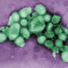
Worldwide, there are about a billion cases of flu every year. Most of them occur during the winter in each geography, with the virus making a huge continuous lap of the globe and evolving as it goes. It’s this gentle genetic “drift” that means the flu can always present a fresh face to our immune systems each year, and keep on infecting us throughout our lives. We combat this by periodically updating our vaccines to better reflect the circulating strains. Traditionally, scientists have gauged the likely effectiveness of those vaccines by looking at the antibodies they provoke; they also keep tabs on the underlying genetic changes that the virus is accruing. But which, if any, is the better guide, and can we improve on it, perhaps by considering both elements side by side? Speaking with Chris Smith, Amanda Perofsky is at the University of Washington…
Amanda - So traditionally the antibody data and the genetic sequences have been analysed separately, just totally in isolation. Scientists look at genetic mutations in the flu viruses and then separately they look at data from the antibody experiments. But now there's more recent methods where we can combine both of those two different sources of information on flu evolution into one analysis. And the analysis is that we create family trees with the flu viruses that aren't just informed by the genetic sequence data, but also by the antibody data.
Chris - And how do you then work out how severe an impact that particular combination will have?
Amanda - So we have historical data on flu outbreaks in the US, and this data is publicly available provided by the US Centers for Disease Control and Prevention. And so there's about 20 years of historical data on people going to the doctor for flu-like symptoms and how those doctor's visits for flu-like symptoms compare to sort of like the overall rate of people going to the doctor. So these are the number of people going to the doctor for flu versus everyone going to the doctor. We can get a rate of flu-like illness over the years and we can break that down by geography as well. We look at each flu season, what was the size of the epidemic, when did the epidemic peak, how severe was the flu season? There's also data collected on deaths that are potentially attributable, which age groups are being infected more in a particular season. And from those family trees of the flu viruses, we can look at how much has the population of flu viruses as a whole changed compared to the last season. And so we get like a one-to-One comparison of this is how much the flu virus population has evolved compared to different aspects of the flu each season.
Chris - And what does that tell you when you've done that? What trends or what pattern emerges in terms of being able to make a better prediction regarding how, how bad a flu season's likely to be based on what the virus is doing?
Amanda - So what we found matches up with the long-held expectations concerning evolution of the flu viruses causing people to become more susceptible to the flu. And in that I mean that the viruses are so different than what you've seen your body or your immune system has seen before, that they're able to infect you even though you've been infected in the past or vaccinated in the past. And so we found further evidence that when flu viruses evolve, that causes bigger flu epidemics. But a thing that was unique about our study is that we looked at all sorts of different ways to measure flu evolution. I mentioned that you can measure flu evolution using the antibody data or the genetic data. What we found that was sort of surprising was that changes in flu genetic sequences were actually more predictive of different aspects of flu epidemics than the antibody data. Even though the antibody data has been considered the gold standard for measuring changes in flu evolution over time
Chris - Is the implication of that then that what we should be doing in future is when we collect samples of flu from patients, we should be looking at how it is evolving and using that evolutionary data to make predictions about vaccine effectiveness about the scale of a likely forthcoming outbreak, rather than assuming, oh look, we've got a reasonable match between the circulating virus and what's in the vaccine, so we're probably gonna be okay...
Amanda - Scientists are already looking at changes in genetic sequences. It's just that perhaps the genetic sequence data should be considered at the same level this gold standard data has been considered in, in terms of determining how much flu viruses have evolved since the last season. Another finding I didn't mention is that traditionally scientists have looked at evolution in one particular gene of the flu virus that is considered to drive the majority of the flu virus escaping detection by immune system. But there's another gene as well we looked at that isn't currently really considered in surveillance of flu evolution. And so we found that this other gene also has just as strong of a relationship with different aspects of flu outbreaks as the gene that scientists have traditionally looked at in the past.

28:51 - How does aphantasia affect autobiographical memory?
How does aphantasia affect autobiographical memory?
Cornelia McCormick, University Medical School, Bonn
Close your eyes and picture what you were doing at breakfast time today. Most people can do that in, literally, the blink of an eye. We can summon up an autobiographical narrative of where we were, what we were doing, who we were with, and the order in which those things occurred. They accompanied by rich visual pictures of the events. But some people can’t do this. It’s a condition called aphantasia, and Cornelia McCormick, from University Medical School, Bonn, has a track record in studying it. As she explains to Chris Smith, she was wondering how being aphantasic affects a person’s memory, and what differences brain scans might reveal when individuals with the condition try to recall things about their lives…
Cornelia - Most of us, if we think about important events of our lives, our marriage, when my children were born, I have very vivid mental imagery in my mind's eye, and in the memory research community there's this understanding that those vivid imagery are really important for autobiographical memories. And one of the questions we have, if the images are missing, can we still remember and what do we then remember of our own lives? And that's when we turned to aphantasia together with the aphantasia research project of the University of Bonn, Merlin Monzel. We got talking and he said, well, why don't we sit together and work on how aphantasics - so people that don't have mental imagery, don't have the images in their mind's eye - then remember autobiographical memory.
Chris - What was the next step then? So having decided this was going to be the group to study who could help you answer that question, how did you put the study together? What were the questions you were trying to ask and how?
Cornelia - Our study had two important parts. One is an interview of autobiographical memories. People are asked to explain in much detail five autobiographical memories. So which detail comes to mind when you remember important days of your life. And then we put people in the MRI scanner and here we were particularly interested in which parts of their brain are activated when they remember their memories.
Chris - Presumably you are comparing people who do and don't have aphantasia and you can then ask, well, which bits of the brain are connected together? Are there any differences in these connections which are flashing up?
Cornelia - Yeah. So we had very strong hypothesis about which brain regions may be involved in this task. We thought the hippocampus is really important because the hippocampus has a longstanding history of being the memory hub of our brain. And the other important brain region is the visual perceptual cortex. So there at the occipital lobe where we process vision in our brains and those two brain regions, hippocampus and occipital lobe, those were the key areas we wanted to investigate. And hypothesised that there are differences between people with aphantasia and people who have mental images in their mind's eye.
Chris - Well, let's begin with what we would dub normal, the people who do have the mental images in their mind eye first. When you do these functional imaging studies, what flashes up in those regions you're interested in, in those normal people?
Cornelia - Well then we can see a strong hippocampal activation and also activation in the occipital cortex. There are also other areas of the brain activated when you remember autobiographic memories. But these were the areas that we were specifically interested in.
Chris - And so a loose interpretation of that is that when you are remembering things and presenting them to your consciousness, because it's got a visual element, the visual system flashes up. Would that be an initial interpretation?
Cornelia - Yeah, so the theory behind it is that the visual or the occipital cortex is providing all those vivid mental imagery details that you need to actually see something in your mind's eye. And the hippocampus is more the hub connecting memories and also providing and constructing a mental scene in your mind's eye.
Chris - And so one would hypothesise then that people with aphantasia who don't see the images might try to compile an experience through their hippocampus and recall a memory, but they wouldn't have any visual activation to the same extent because they're not seeing anything in their mind's eye?
Cornelia - Exactly. So now we are going to the really exciting results. So I would like to just go back to the interview one second because that's already very interesting what happens in the reports of their memories. So here we find that not only are the visual details missing to the autobiographic memory, the whole memory system seems to be impacted. So they also have hardly any emotional details to their memory and they had a hard time remembering memories at all. So it's not only that the visual details are missing, but the whole autobiographical memory seems to be deficient in a way.
Chris - And if you compare how strongly the different parts of the brain are being activated in the two groups, because if they're trying harder to try to recall memories because something's amiss, you might see a disparity. So how does the activation that you do see compare between the two groups?
Cornelia - Exactly. So we found that the visual perceptual cortex or the occipital cortex actually had more activation for people with aphantasia than for our control participants. And the hippocampus, on the other side, had less activation in people with aphantasia than in controls. And we thought that the hippocampus is impacted because the entire memories are deficient. It's not only the visual part, but also the global autobiographical memory. And for the visual cortex, at first we were a bit surprised about that because we thought maybe because they don't have the images, also the visual perceptual cortex would also have less activation. But it's actually known in the literature that the visual perceptual cortex has more activation in aphantasia. And this may be a cause of aphantasia. There are theories that the visual cortex has too much activation in aphantasia and all the small signals that you need to see mental images in your mind's eye, which are not so stark as the outside world don't exceed the activation in the visual cortex.
Chris - It's almost like not being able to see the wood for the trees: you've got so much noise going on that you end up recruiting a whole noisy milieu of messages, which, effectively, confuses the experience. So you don't actually form a coherent mental image?
Cornelia - Exactly. That's what we think. We don't know for sure yet, but that's how we interpreted our results.
Chris - Fascinating. What happens then if you do the same study on people blind since birth?
Cornelia - Also very interesting question because we are just acquiring those data and we haven't analysed the data yet, so I don't know yet. But it's a fascinating research question!
Chris - There has to be something more to this though, doesn't there? Because people who have been blind since birth have excellent memories. So it's not just a question of being able to see things that make you very good at remembering things, is the message that I would take away from that?
Cornelia - That's what I think too, although there's very, very little research on this done with like state of the art autobiographic memory interviews and also MRIs. So that's why we are doing this kind of more rigorous study and experiment to see how they compare with sighted people, right. Maybe, maybe their memory just works differently because, I agree, if you're blind, you have to have a good memory because otherwise if you put your cup of coffee somewhere else and you can't remember where you put it, you don't see it, right, so you have to have this memory to relying on finding your object in your environment again.
Related Content
- Previous ADHD explained
- Next Why do we dream the things we dream?


Comments
Add a comment