In this NewsFlash, we find out how lasers can cut complications in cataract surgery, why some people are allergic to wine and discover how scientists have solved a long-standing mystery relating the structure of the placenta. Plus, we get a rundown of the highlights from the world's biggest neuroscience meeting, Neuroscience 2010 in San Diego.
In this episode

00:25 - Laser cataract surgery a sight for sore eyes
Laser cataract surgery a sight for sore eyes
One of the world's most commonly performed surgical procedures - cataract surgery - could soon be revolutionised with a laser. Stanford University scientist Daniel Palanker and his colleagues, writing in Science Translational Medicine, have developed a laser mapping and cutting technique to significantly improve the safety and success of lens replacement surgery, which up to one third of the developed world's population undergo  Rakesh Ahuja, MD") in their lifetimes.
in their lifetimes.
Cataracts occur when the lens, which sits at the front of the eye behind the cornea, becomes cloudy; this hinders the passage of light into the eye, impairing visual acuity, colour perception and making it especially difficult to see in low light conditions. Ophthalmic surgeons remedy the situation by first making an incision in the front part of the eye. They then painstakingly perform a technique called a capsulorhexis, which involves opening up a circular aperture in the capsular membrane that holds the damaged lens. This incision must be circular to avoid producing any "edges" that would be weak spots prone to rupture. When this is complete the lens is broken up, usually by ultrasound, and removed through the aperture. A replacement prosthetic lens is then inserted in its place. Although the the operation has an very high success rate, there are complications.
The trickiest step is performing the capsulorhexis, which relies entirely on the skill of the surgeon and his or her estimation of the "correct" placement of the incisions during the procedure. To remove this uncertainty, the Stanford team have designed a femtosecond laser-assisted technique that initially maps out the eye, performs the capsulorhexis procedure and also cuts up the lens into tiny fragments facilitating its removal. Using pig's eyes to hone the technique and then rabbits to test its safety, the new approach produced capsulorhexis results that were twice as strong as those performed free-hand by a surgeon, thus potentially reducing the risk of complications. The system was then tested on 50 patients - though not as a blind trial (!) - 30 of them in a cross-over study whereby one eye was treated via the traditional surgical technique, the other using the laser.
There were no negative outcomes, confirming the safety and effectiveness of the approach, the cuts made by laser were 50 times more precise than those made by hand and, although stastically not significant, the laser-treated individuals also had slightly better visual acuity afterwards.

04:08 - Why some people are allergic to wine
Why some people are allergic to wine
A lot of us like a nice glass of wine at the end of the day, but for around 500 million people in the world, that's not so much fun, as they have an allergy to wine. It's apparently a bit like having a hayfever attack, with itchy eyes and sneezing. Now a group from the University of Southern Denmark have found out exactly what it is in the wine that makes these people react to it.
It's a type of molecule called a glycoprotein, which is also what causes other allergic reactions like those to pollen and dust mites. These are molecules made up of sugar and protein molecules joined together and they're really important to life - they're found in our cell membranes, some important hormones are glycoproteins, and they also play a key role in immune response. The kind of glycoproteins on the outside of an invading pathogen like a bacterium for example help the immune system to identify it.  rosevita (Morguefile)")
So the team led by Guiseppe Palmisano tested an italian chardonnay using a mass spectrometer to see if there were glycoproteins present that might be causing these reactions. They identified 28 different glycoproteins in the wine, contributed by both the grapes and the yeast used to make the wine. Many of the glycoproteins from the grapes were very similar in structure to ones known from other plants and fruit that are known to cause allergic reactions, and it's the structure of the glycoprotein that is important in it being an allergen or not.
So the team suggested that this could be a step towards creating low-allergenic wines, although this would probably be a fair way off as the very glycoproteins that can cause these reactions are essential for the palate and the structure of the wine, and it's not clear exactly which of the glycoproteins present might be responsible, so there's still work to be done there...
06:14 - Highlights from the Society for Neuroscience Meeting 2010
Highlights from the Society for Neuroscience Meeting 2010
Smitha Mundasad
Chris - Also this week, the annual meeting of the Society for Neuroscience took place in San Diego, California; a nice sunny place. This is actually the largest meeting of neuroscientists from around the world who meet to discuss their latest research and the progress they're making in understanding how the brain works. Naked Scientist Smitha Mundasad was there for all the action and she's with us now to tell us what she got up to. Hello, Smitha.
Smitha - Hi, Chris.
Chris - So the weather here, decidedly worse than California then?
Smitha - It's so much nicer in California.
Chris - So, a shame to be back, but what did you get up to?
 Dwayne Reed @ Wikipedia") Smitha - Well, with over 30,000 neuroscientists in attendance, this year's Society for Neuroscience Conference in San Diego, was a hot bed of new research and exciting ideas. One of the meeting's highlights was news that researchers have found a way for people to control computer cursors with their thoughts alone. Using MRI machines connected to computers, scientists from the University of Pennsylvania School of Medicine had 14 participants think alternatively of two thoughts: One, to think about playing tennis; and the other, to imagine going from room to room in a familiar place. Analysing the brain activity from these two different scenarios, the researchers were able to show that the computers could distinguish two distinct patterns of brain activity for each thought. While still in the MRI scanner, the participants were asked to use these thoughts to control the movement of a computer cursor on a screen. This means that they had near instant feedback of how well they could control their own thoughts. Lead author, Dr. Anna Rose Childress explains that how new approach could have major therapeutic implications, for example, in the treatment of addiction...
Smitha - Well, with over 30,000 neuroscientists in attendance, this year's Society for Neuroscience Conference in San Diego, was a hot bed of new research and exciting ideas. One of the meeting's highlights was news that researchers have found a way for people to control computer cursors with their thoughts alone. Using MRI machines connected to computers, scientists from the University of Pennsylvania School of Medicine had 14 participants think alternatively of two thoughts: One, to think about playing tennis; and the other, to imagine going from room to room in a familiar place. Analysing the brain activity from these two different scenarios, the researchers were able to show that the computers could distinguish two distinct patterns of brain activity for each thought. While still in the MRI scanner, the participants were asked to use these thoughts to control the movement of a computer cursor on a screen. This means that they had near instant feedback of how well they could control their own thoughts. Lead author, Dr. Anna Rose Childress explains that how new approach could have major therapeutic implications, for example, in the treatment of addiction...
Anna - Control of the screen cursor is a really good measure of how well a person can alternate their thoughts, between tennis and room by room. The thoughts, of course, are completely arbitrary, but the act of controlling them and shifting them does require considerable attention and cognitive control. For our patients, when they are in the real world and maintaining cognitive control while they're driving their car down the street in a cocaine neighbourhood, what they describe is that they will be intruded on by a brief vision of something cocaine related and they become derailed. So we're going to be able to model that with this task. We'll have people performing this task and be blipping in very brief cocaine images and be able to actually see the brain struggling to maintain control. So it's a very sensitive probe for disrupted cognitive control in pathological conditions such as addiction for example.
Smitha - Anna Rose Childress from the University of Pennsylvania.
In other news from the conference, again combining the disciplines of computing and neuroscience, researchers have found a novel way to make blind mice see. There's over 25 million people worldwide who suffer from degenerative diseases of the retina, often resulting in partial sight and blindness. Photoreceptors on the retina normally receive light and then, with the help of retinal ganglion cells, this is converted into electrical impulses that can be understood by the brain. But with many retinal diseases, these photoreceptors stop working.
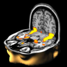
, on the ventral bank of the calcarine sulcus (check this last point). The brain's surface is extracted from structural MRI data (Wellcome Dept. Imaging Neuroscience, UCL, UK). © Original uploader was Washington irving @ wikipedia") Existing retinal prosthesis offer very limited hope. Implanting electrodes into the retinal cells can allow people to make out spots of light or edges of objects, but very little in the way of real vision.
Existing retinal prosthesis offer very limited hope. Implanting electrodes into the retinal cells can allow people to make out spots of light or edges of objects, but very little in the way of real vision.
But now, researchers at Weill Cornell Medical College in New York have taken a new approach. By analysing the light input and then the corresponding neural output of the healthy mice retinal cells, they were actually able to mimic the way the retina converts light into electrical signals. They've actually essentially cracked the neural code used by the brain. This code can then be used to produce a much sharper, clearer image in mouse models. The team hope to work with primates next and then humans very soon. Lead author, Sheila Nirenberg, explains how her research differs from other approaches...
Sheila - Our common analogy is that the patient's eye is like a digital camera with damaged pixels. So the more of the pixels you replace, the better the picture you're going to get. What our research shows is that there's another factor that's just as critical. Not only do you need to stimulate large numbers of cells, but you also have to stimulate them with the code that the eye is sending to the brain. This is because the camera analogy really only holds in part. The eye does essentially take a picture, but then it goes much further. It processes the picture. It extracts information from it and then it converts that information into a code that the brain can read. So, to make an effective prosthetic device, you've really got to have both these functions: acquiring the picture and then converting the picture into a code that the brain can make use of, and I think we really have this now. We have both of these components.
Smitha - Sheila Nirenberg from Weill Cornell Medical College, New York.
There was also good news for musicians at the Society for Neuroscience Conference. Benjamin Zendel of the University of Toronto presented research that suggests that musicians may actually be protected from some of the age-related changes in the auditory cortex of the brain. The researchers presented participants with complex sounds under two conditions: One, where they were distracted by another activity and the other, where they were focused on the sounds. During these experiments, the participant's brain activity was measured using EEG. The brain activity patterns of older people with musical training were very similar to that of young people during the attentive listening task, but older non-musicians showed typical age-related changes. Lead author, Benjamin Zendel...
Benjamin - A lot of research has shown that musicians do better on many hearing tasks. They have more acute hearing, they're better at making fine distinctions between sounds, and also the exact same things that change with age. So as you get older, it's harder to make these fine distinctions between sound and that contributes to difficulty understanding speech in noisy environments like at a noisy restaurant or a noisy coffee shop, and so, the really exciting part of this research is that older musicians seem to maintain some of those abilities and it's reflected in changes in the auditory cortex. That there are changes in the functional components of the auditory cortex in older musicians that make their brains effectively look like that of the younger adults.
Smitha - So those piano lessons might have been a bit more valuable than I thought! That was Benjamin Zendel from the University of Toronto.

12:51 - Bacteria cause colour change in aphids
Bacteria cause colour change in aphids
This week, researchers in Japan have shown that the colour of pea aphids can be changed by bacteria living inside them.
The reason this is such an interesting discovery is that colour is a really important aspect of an animal's life. It can influence predators, prey and potential mates.
The team, led by Tsutomu Tsuchida studied these pea aphids, which are from France, and are found in both red and green morphs in the wild. Red ones tend to get eaten more by ladybirds, but the green ones tend to be attacked by parasitoid wasps. So when the team were looking at the wild populations, they noticed that some of the green aphids were having red offspring, but that the red offspring gradually became green as they aged. They wondered what might be causing the colour change, so took a closer look at the aphids and found several different types of endosymbiotic bacteria were living in them.
 Fir0002") An endosymbiont is an organism that lives inside another organism, but the interaction between them benefits both parties - it has to, otherwise it would be considered to be a parasite or an infection. One example would be corals - they have tiny algae living within their cells that photosynthesise - the corals gain energy from their symbionts, and the algae have ready access to nutrients and safety from predators.
An endosymbiont is an organism that lives inside another organism, but the interaction between them benefits both parties - it has to, otherwise it would be considered to be a parasite or an infection. One example would be corals - they have tiny algae living within their cells that photosynthesise - the corals gain energy from their symbionts, and the algae have ready access to nutrients and safety from predators.
So, to find out if the endosymbiotic bacteria in the aphids were involved in the colour changing, the team treated groups of aphids with antibiotics to knock out some of the bacteria. And in a second experiment they injected uninfected aphids with haemolymph, which is essentially like the blood, from infected aphids.
They found that one of the groups of bacteria, called Rickettsiella, was responsible for the colour change.When the other bacteria were killed off using antibiotics, the aphids' colour still changed if Rickettsiella was present, and when it was injected into uninfected red aphids, it caused their red offspring to become green as they aged. When the red aphids produced their sex cells, the bacteria hitched a ride in those cells and were then present in the offspring, making them change colour.The group suggested that this colour change may protect them from predation by ladybirds, but that Rickettsiella is also usually found with two other symbiont infections that help protect against the parasitoid wasps, so helping to offset the danger there of being green.
So it's an example of some of the really complex relationships that there are between insects and bacteria. I mean these ones do convey an advantage by being involved in changing the colour of the aphids, but there are some that are even more bizarre, like Wolbachia, which can cause sex changes, kill off all the males in a brood of insect babies and is estimated to infect up to 70% of all insect species.

15:37 - Plugging into the placenta
Plugging into the placenta
Scientists have solved a long-standing mystery relating the structure of the placenta, the lifeline that connects mother and baby during embryonic development. For over 100 years anatomists have been scratching their heads trying to explain why, despite the function being the same for every animal, the placentae of different species were so dramatically different in structure.
Now two Durham University scientists, Isabella Capellini and Robert Barton, writing in the journal The American Naturalist, think they know why. Their study compared 109 different animal species, including humans, taking into account the size and length of gestation of each animal as well as the stuctures of their corresponding placentae. What Asturnut on wikipedia") they found is that the simpler the structure of the placenta, the longer the gestation. So animals like a human or an ape, which have relatively simple placentae comprising just "fingers" of foetal tissue projecting into the wall of the uterus to contact maternal blood, tend to have a longer gestation, while species like dogs and leopards, which have relatively short gestation times, have extremely complex and highly folded "labythine" placental structures.
they found is that the simpler the structure of the placenta, the longer the gestation. So animals like a human or an ape, which have relatively simple placentae comprising just "fingers" of foetal tissue projecting into the wall of the uterus to contact maternal blood, tend to have a longer gestation, while species like dogs and leopards, which have relatively short gestation times, have extremely complex and highly folded "labythine" placental structures.
This latter configuration provides a very high surface area through which nutrients can be picked up by the developing foetus, while the simpler stucture adopted by humans have a lower surface area and hence a more limited rate of nutrient provision to a baby, prolonging the gestation. Why this is important is that it reflects the metabolic cost of pregnancy and is probably a protective mechanism by which a mother controls the resource conflict between her needs and those of the developing baby. Now this relationship is understood, Robert Barton points out, "it will be interesting to track down the genes involved and this will inevitably have clinical consequences too, informing our understanding of pregnancy-related conditions like pre-eclampsia."


 crystal")

Comments
Add a comment